
This digitally coloured scanning electron microscopic image depicts red blood cells intertwined into the rapidly forming fibrin net.

This coloured electron microscopic picture of a murine kidney shows part of the Bowman capsule (or glomerulus) located in the nephron, where intertwined vessels filtrate the blood fluids to form urine.

This digitally coloured scanning electron microscopic image depicts murine lens cells. The cells of the lens are near to transparent and contain only a small amount of organelles. The entirety of lens cells refract the light in a way to focus onto the retina.

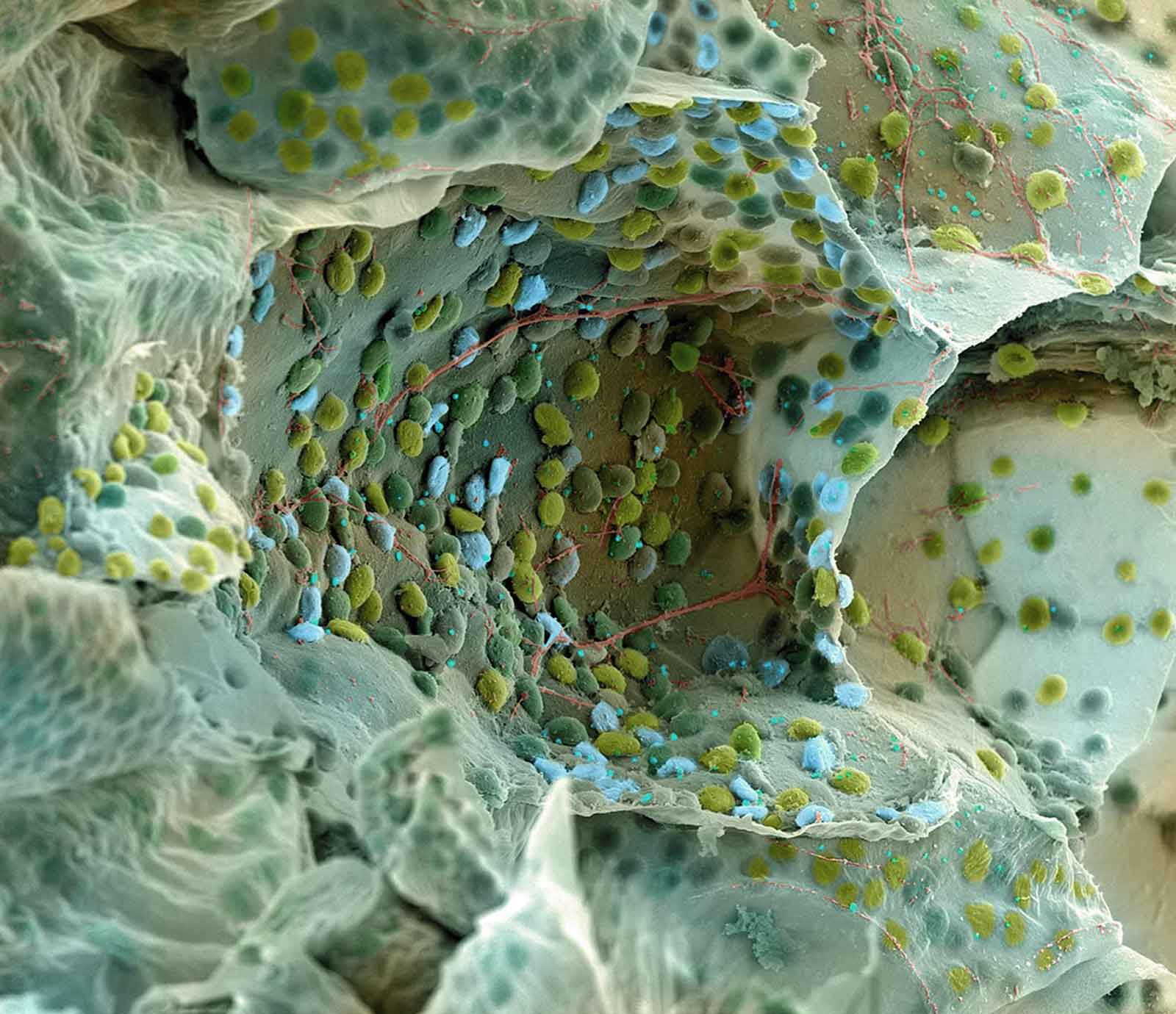
This section of Delospermum cooperi shows the vast amount of chloroplasts lining the internal membranes of the cells.
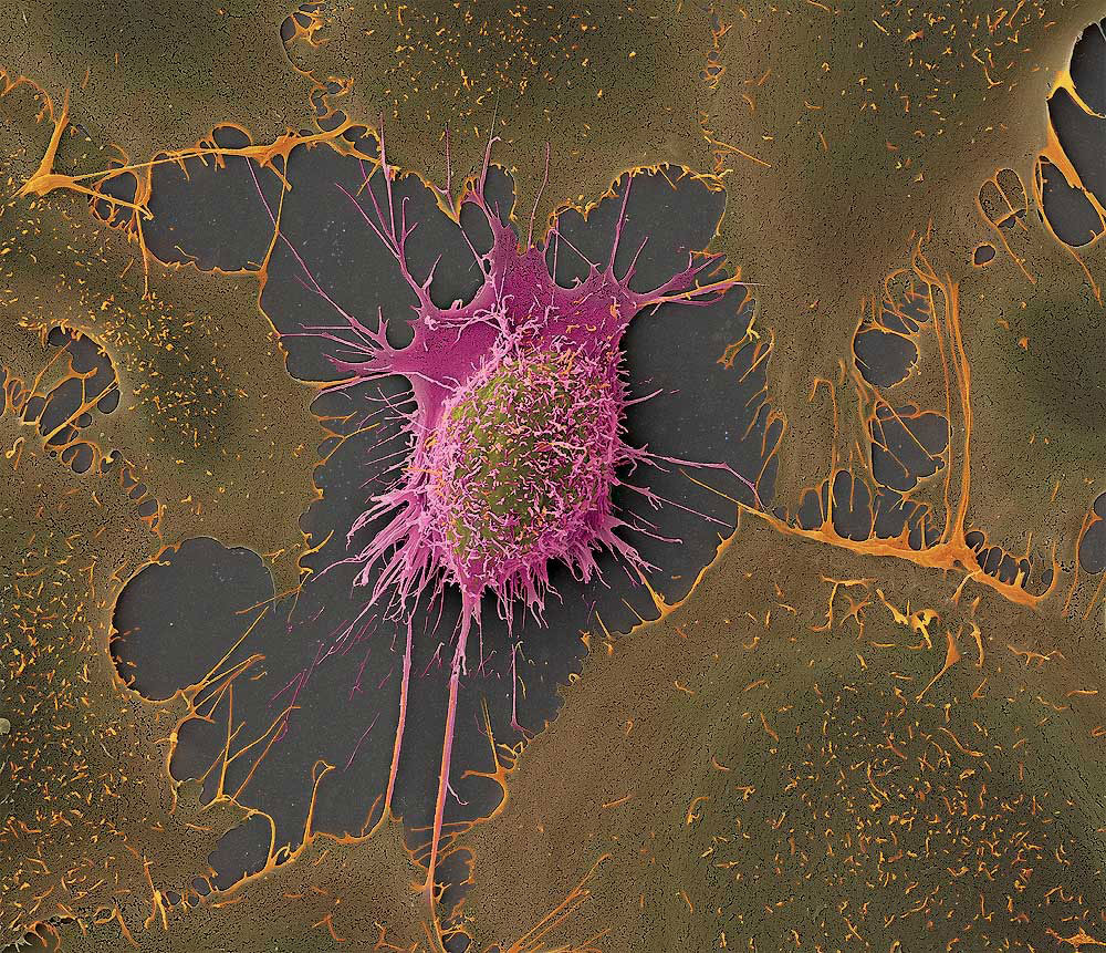
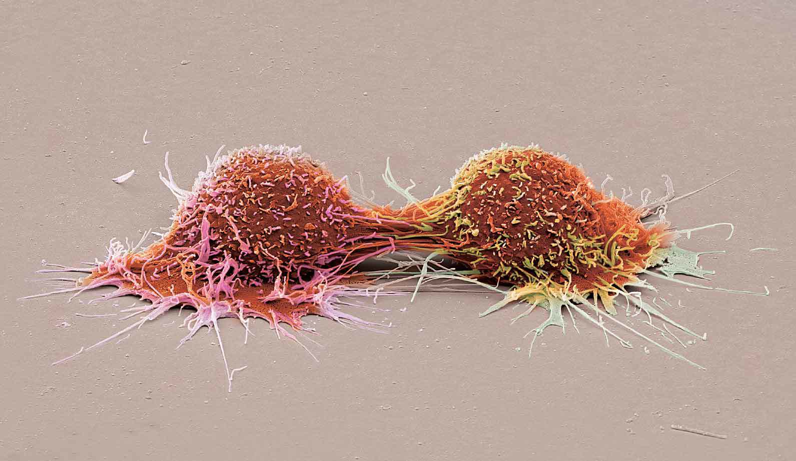
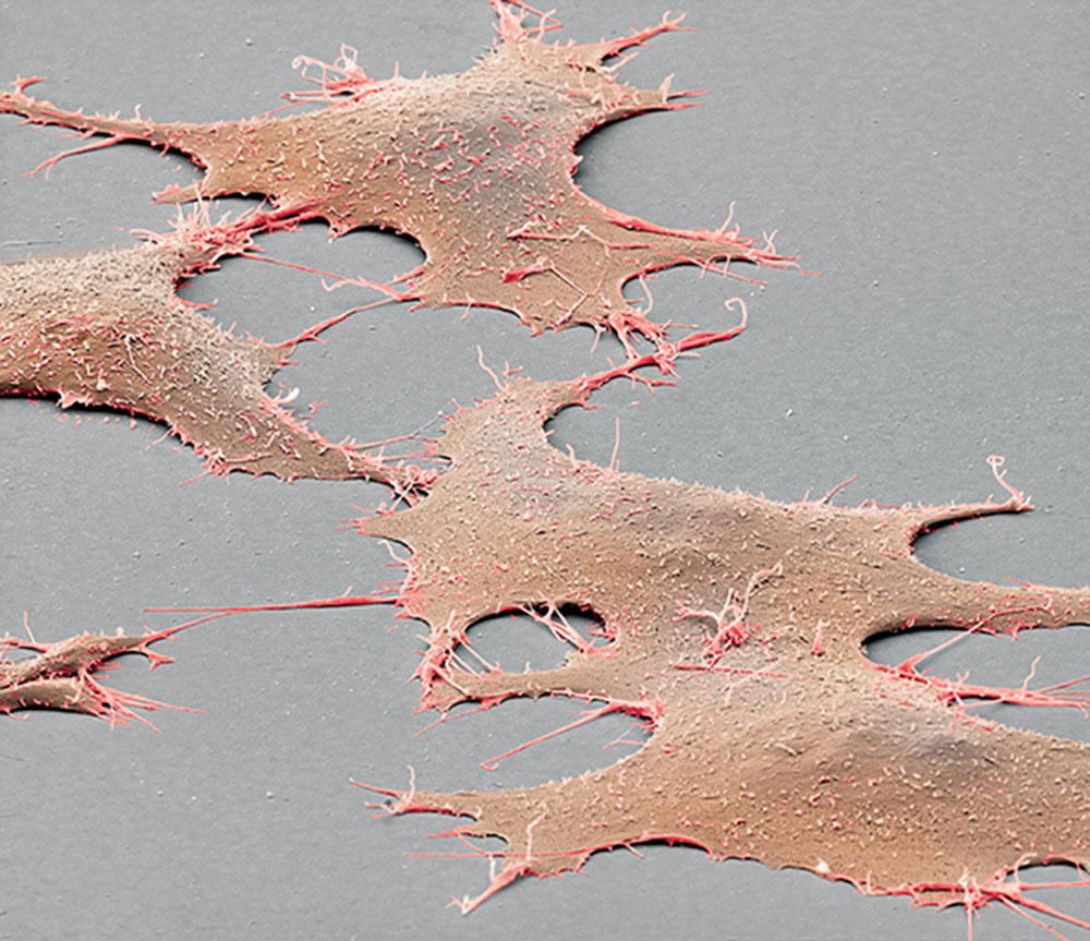
Depicted are cells from a human hepatocellular carcinoma (HepG2). A tumour cell line established from a 15 year old Caucasian boy. These cells have 55 chromosomes instead of 46 as in healthy humans. Cancer cells lose the ability for correct program of dying and consequently proliferate uncontrolled. This fact can be used by researchers not only to study cancer itself, but to use them as a study model, which proliferates indefinitely. In contrast primary established cells from tissue normally die off after a certain number of divisions.
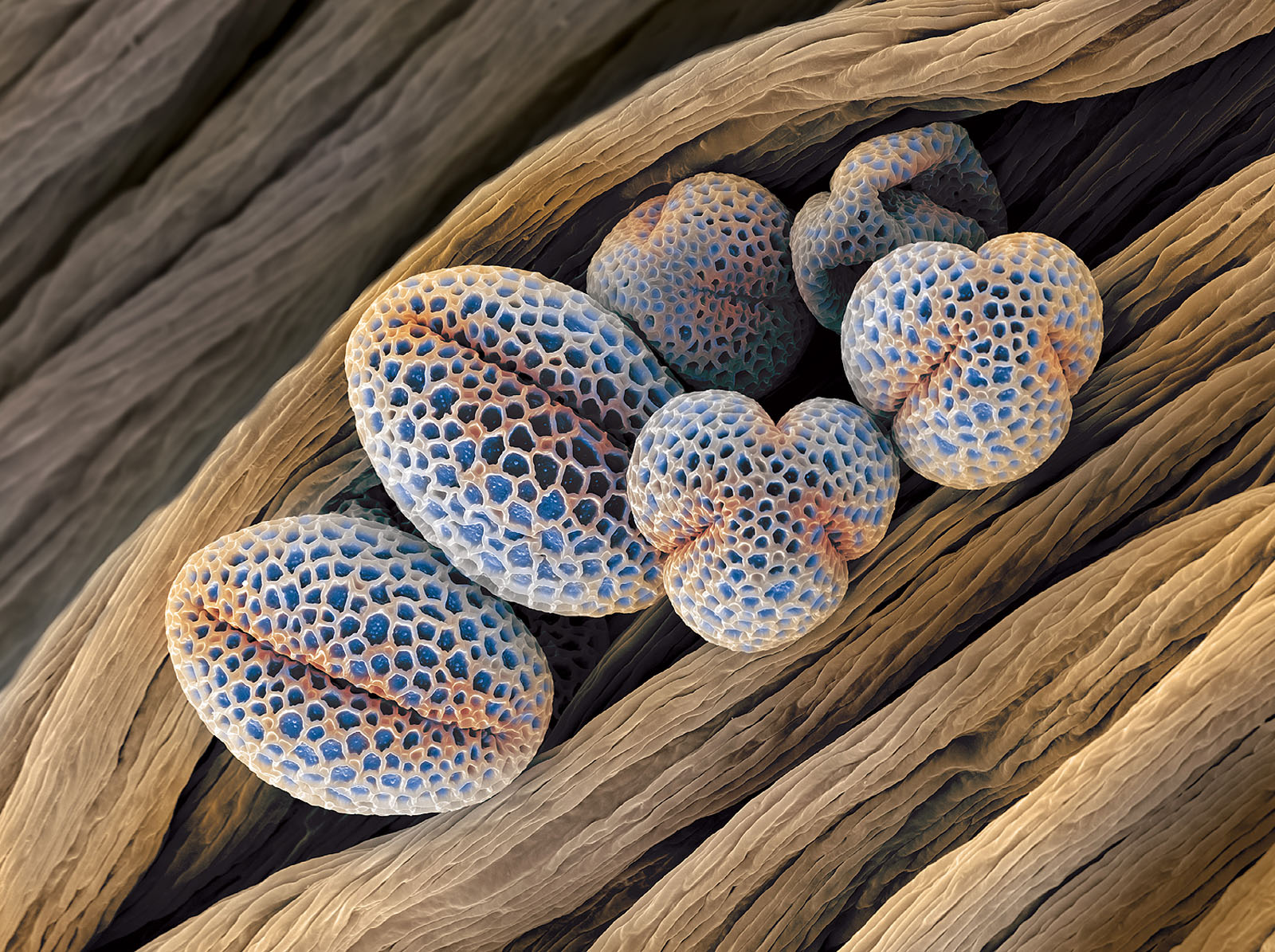
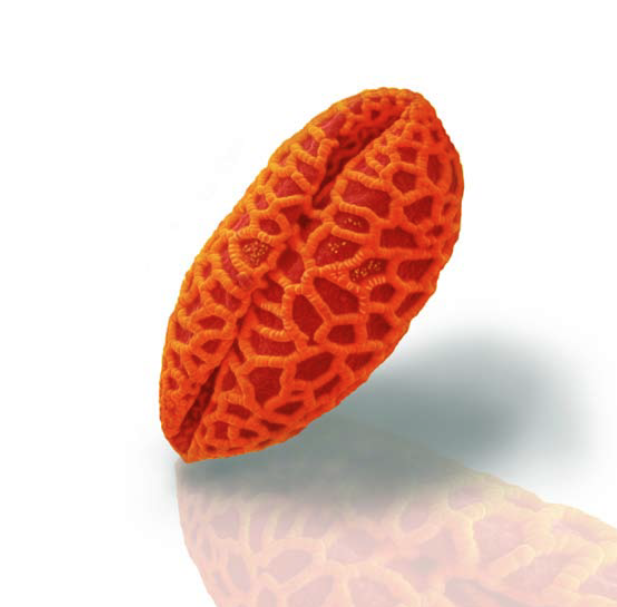
Be amazed - not allergic! Digitally coloured scanning electron microscopic image of Forsythia pollen.

This image depicts cilia in the mouse brain together with a few erythrocytes.
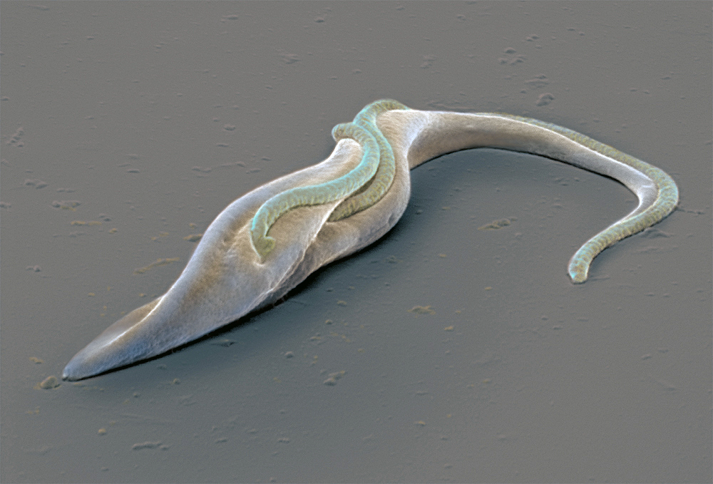
The sleeping sickness in humans and the nagana in animals are caused by the unicellular parasite Trypanosoma brucei brucei. The locomotion of an individual also in the process of division is captured.
Parasite was a kind gift of Prof. A. Schneider, Departement of Chemistry & Biochemistry, University of Berne

The parasitic wasp Eupelmus vuilleti
(Hymenoptera: Eupelmidae). Parasitic wasp mostly use spiders, butterfly
or bug larvae as host. Every species is specialised mostly only one
host. Eupelmus v. in his right is a solitary ectoparasite of the larvae of the bean weevil (Acanthoscelides obtectus). Although small (2-3 mm) Acanthoscelides o. can destroy entire seed stock.
Specimen was a kind gift of Dr. C. Lüthi
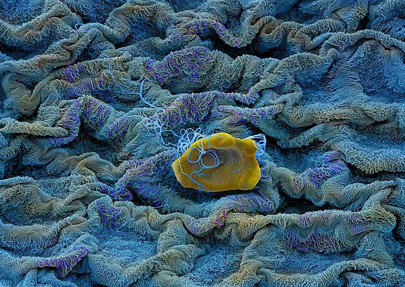
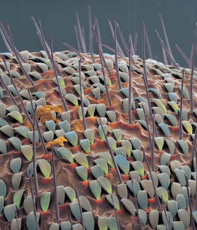

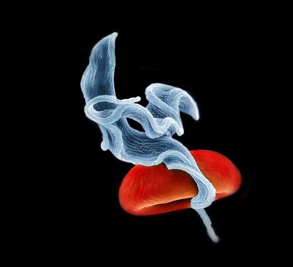
This image depicts two Trypanosomes - causative pathogen of the sleeping sickness in humans or Nagana in cattle and one red blood cell. Its vector is the TseTse fly.
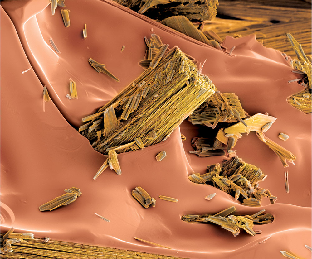
Yes, the very thing you drink in the morning.
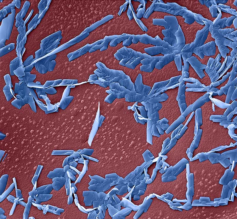
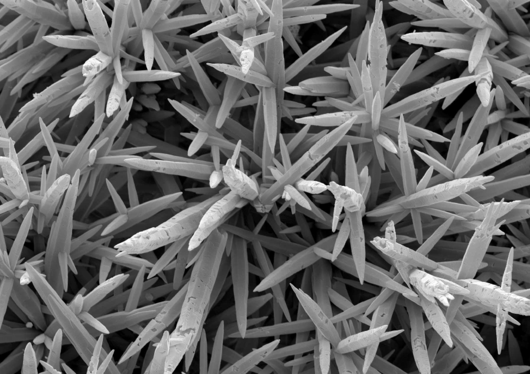
This coloured scanning electron microscopic picture depicts a crystallised compound - nucleotide -, which makes up the building blocks of the DNA.
